
Visual signs of disease can often be spotted within affected tissues, and advances in histopathology have provided clinicians with powerful diagnostic tools to spot those signs. Microscopes are the cornerstone of this trade, and although they have proven to be extremely useful, they do suffer from some limitations. They are effectively 2D imaging devices that don’t offer a good perspective on the volumetric nature of things at small scales, and they only detect light, which can’t sense clinically important properties such as tissue stiffness.
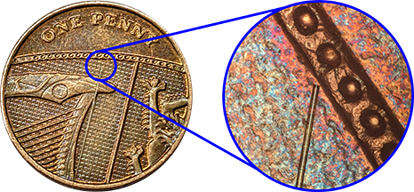
Now, researchers at University of Nottingham in England have developed a nanoscopic ultrasonic imaging system that can be coupled with conventional optical devices to visualize cells and their components in a new way. The imaging probe is so small that it can barely be seen on a small coin.
Unlike light, ultrasonic imaging can provide nuance about the 3D structure of whatever is observed. Moreover, since sound generates waves within the tissues it comes in contact with, sensing the properties of those waves can offer a lot of information about the nature of the tissues themselves. “We believe the system’s ability to measure the stiffness of a specimen, its bio-compatibility, and its endoscopic-potential, all while accessing the nanoscale, are what set it apart. These features set the technology up for future measurements inside the body; towards the ultimate goal of minimally invasive point-of-care diagnostics,” said Dr Salvatore La Cavera III, one of the team leaders that developed the new device.
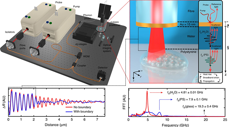
The tiny size of the new probe allows it to be used alongside optical components of conventional endoscopes, and the two imaging modalities can be combined to provide a more comprehensive assessment of tissue. As the photons of light bouncing back from tissue are represented as bright points on a display, so are the phonons from the ultrasound probe. Because the technology fits the tips of optical fibers, it can be used to visualize difficult to reach targets such as those within the GI tract.
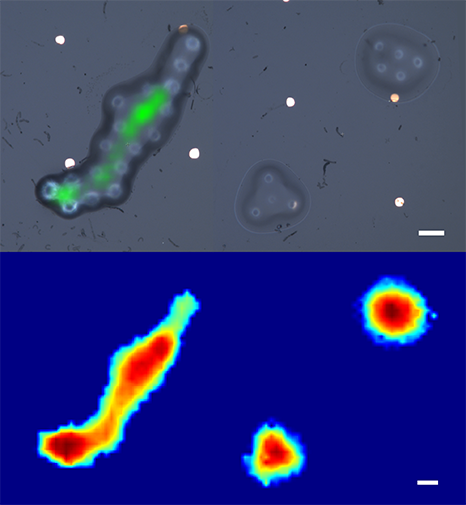
The ultrasonic probe has an unusual characteristic that allows it to image in 2D at microscopic scale, but in the 3rd dimension it images at a much more impressive nanoscale.
Some details about how the new device works, according to University of Nottingham:
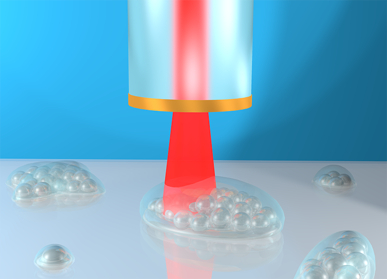
The new ultrasonic imaging system uses two lasers that emit short pulses of energy to stimulate and detect vibrations in a specimen. One of the laser pulses is absorbed by a layer of metal – a nano-transducer (which works by converting energy from one form to another) – fabricated on the tip of the fiber; a process which results in high-frequency phonons (sound particles) getting pumped into the specimen. Then a second laser pulse collides with the sound waves, a process known as Brillouin scattering. By detecting these “collided” laser pulses, the shape of the traveling sound wave can be recreated and displayed visually.
The detected sound wave encodes information about the stiffness of a material, and even its geometry. The Nottingham team was the first to demonstrate this dual-capability using pulsed lasers and optical fibers.
Open access study in Nature journal Light: Science & Applications: Phonon imaging in 3D with a fibre probe